Foreign bodies occur when your pet eats an object that is unable to pass through the gastrointestinal tract easily. Examples of this can include; household objects, toys, bones and certain foods that can’t be digested. These objects can cause problems such as obstruction of the gastrointestinal tract, damage and perforation of the intestines and toxicity from certain materials, such as metal. The severity of the problems caused depends on the duration the foreign body has been present, the location of it, the degree of obstruction it has caused and any problems associated with the material of the object. If perforation of the intestinal tract occurs and the contents leaks into the abdomen this can quickly lead to inflammation of the abdominal lining (peritonitis) and allows bacterial contamination (sepsis), both of which are life-threatening conditions.
If you see your pet eat something that could get stuck in their intestinal system, we would recommend you phone the practice as soon as possible. If we see an animal soon after they have ingested an object sometimes it is appropriate to induce vomiting to try and bring it back up. This is only an option shortly after the object has been swallowed because once it passes the stomach it cannot go back. We would not induce vomiting if the object could cause more harm on the way back up the oesophagus.
The common clinical signs of foreign bodies include:
- Vomiting
- Anorexia (loss of appetite)
- Abdominal pain
- Depression
- Dehydration
- Diarrhoea
These signs can vary depending on the severity of the foreign body. They can also be seen with other types of gastrointestinal disease, for example dietary indiscretions or viral infections therefore your pet will need a thorough physical examination to help differentiate between these diagnoses. If these signs persist for a prolonged amount of time they themselves can cause further problems too so an appointment with your vet is important. Lots of things will influence what the vet will decide to do next, this includes the animals age, breed, clinical history and physical exam. If they are suspicious that your pet has a foreign body then imaging diagnostics such as ultrasound and radiographs are often helpful in the diagnosis of one.
Ultrasound images don’t often show the actual foreign body but they can show the effects the obstruction is causing. For example, a build-up of gas or fluid that cannot pass the object. If the foreign body has damaged part of the gastrointestinal tract and the contents are leaking out this would also show up as abnormal free fluid within the abdomen.
Radiograph images can also show gas or liquid but radio-opaque foreign bodies may also be visible. This means the object absorbs x-rays so can be seen on the image i.e metal materials.
If there is strong evidence of a foreign body, then the vet will decide the most appropriate treatment approach. In many cases this involves surgical exploration of your pets abdomen to find and surgically remove the foreign body. In occasional cases, whereby we think the foreign body may pass through itself then your pet may be given time to allow this to happen, with close monitoring and support to ensure no problems arise.
We have seen a few foreign bodies at Hollybank recently, here are some interesting examples for you!
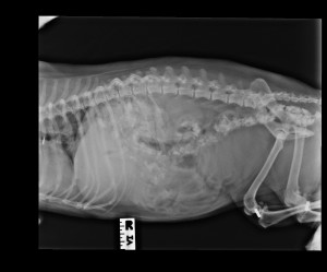
 Cassie’s owner reported seeing her eating from the cats’ litter tray! The x-ray shows a gravel like consistency in the intestines but there was no evidence of an obstruction. An ultrasound scan confirmed Cassie’s guts were moving normally which was good. Gut stasis (reduction in normal movement) can sometimes be an issue with gastrointestinal disease. As the cat litter was so small and had already reached the largest part of the intestine (the colon) it was decided to allow Cassie time to pass these through naturally. She was closely monitored during this time for any changes and kept comfortable. Cassie passed the gravel soon after.
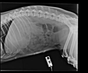
 Pip presented vomiting and with abdominal pain. Suspicious of a foreign body we elected to ultrasound scan her abdomen. This showed lots of gas build up in her small intestine which can be consistent with a gastrointestinal obstruction. We took an abdominal x-ray which too confirmed a gas build up. However, it was not apparent what was causing this obstruction. This is because some materials do not show as well. Although we could not see an obvious object, the combination of Pip’s clinical signs and the suspicious signs on her x-rays gave us enough evidence to warrant surgical investigation. Pip had in fact eaten a corn on the cob! This was removed and she was back to her normal self in no time!
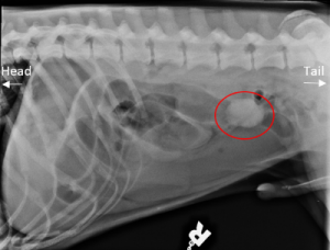
 Roscoe’s x-ray shows a round shaped object in the middle of his abdomen with a build-up of gas in the intestines prior to this. The clinical signs and images were consistent with a suspected foreign body and surgery was performed to remove the object. This one turned out to be a chewed up rubber ball! Roscoe also recovered quickly from surgery.
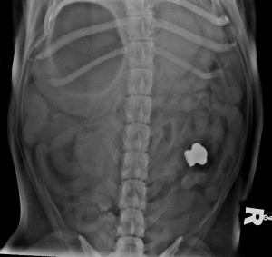
 On Daisy’s radiograph you can clearly see the foreign body as it was a metal material. This foreign body caused Daisy to become unwell and she required surgery to remove it. Her recovery was slightly longer than normal as we suspect there was an element of metal toxicity too. She recovered well and is back to normal.
Fortunately, all of these foreign body patients have made good recoveries and all of our patients needed this vital surgery. However, gastrointestinal surgery is not without its risks and prognosis is improved by identifying the problem and addressing this in a timely manner. If you think your pet may have eaten something or is showing concerning gastrointestinal signs then it is always best to get your pet checked out.


