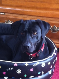
Meg recently came to see us with a lump on her side, just behind her armpit. It was very small, only about 1 cm wide and wasn’t bothering Meg at all. However it had grown slightly over a few months so her owners wanted to get it checked out.

Now dogs and cats can get lots of different types of lumps, with lots of different causes. Unfortunately just by examining a lump it isn’t always apparent what the cause is as many can look the same. There are however a few factors that can give us an idea about whether a lump is benign (doesn’t spread and usually only causes problems due to size and location) or malignant (can spread and cause secondary disease). Usually benign lumps are slow growing, don’t change quickly or cause problems. Contrary to this, malignant masses usually grow more quickly and can causes other problems and signs. However this is not always the case. We have seen several malignant lumps which are very small or have been there for a long time before suddenly causing a problem.
As we were not able to tell what Meg’s lump was we decided to take a sample from it. One if the easiest ways of doing this is to put a fine needle into the lump to remove some cells. These cells can then be smeared onto a slide to be examined at the laboratory. Usually we can take this sample very quickly and easily and it doesn’t require any sedation or an anaesthetic.
Meg’s results came back as a Mast Cell Tumour which is a type of malignant tumour that has the potential to spread and cause further problems. There can be several different types of mast cell tumour, some of which can act more aggressively than others. A higher ‘stage’ of tumour suggests a more aggressive one. From the needle sample we took we were unable to know what stage Meg’s tumour was. Therefore the next step was to remove it and send the whole lump off to the lab for further examination. This was important to find out if further treatment, such as chemotherapy, radiotherapy or further surgery was needed.
Because this type of tumour can release histamine granules and particles into the surrounding skin we gave Meg some antihistamines prior to surgery. We also had to take wide margins of tissue when removing the lump. This was made a little tricky by it’s location just behind her armpit as there isn’t much tissue to spare in this region. In some cases when we struggle to fill the gap we use a skin flap from another area close by. Luckily this wasn’t needed for Meg and her skin closed together really well after we removed the lump.
The lab confirmed that Meg’s Mast cell tumour was an intermediate grade. This meant that surgery was likely to be curative, although it is important to monitor for any signs of recurrence. Surprisingly in the skin we removed Meg also had a second Mast Cell Tumour which was so small it was just a fleck of white on her skin. This Mast Cell Tumour was low grade and may have been unrelated to the first, or some local spread of the first mass.

After the surgery Meg was strictly rested so that her skin wound could heal as easily as possible. She also had some pain relief and antibiotics after her operation. Meg’s wound has been healing well and we have now removed her staples post operatively. Once her fur grows back it won’t be easy to even tell she has had surgery. It was important that Meg’s owners were so observant and noticed Meg’s lump had changed as we were able to remove it early. If you notice any lumps or bumps on your dog or cat, particularly if the appearance has recently changed it is worth getting it checked out by one of the vets. Meg can now continuing enjoying life, especially now she can get out and about again!


