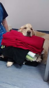
Brave Pet of the Month…Freddie
 Freddie is a gorgeous Golden Retriever who presented to Hollybank with breathing difficulties. Freddie had been very well prior to this at home and was currently in kennels for a short stay with another Golden Retriever, in fact his mum, Lucy.
The kennels had noted Freddie was snuffly, quiet and a little off his food over the last 24 hours. Suddenly that afternoon Freddie collapsed with heavy breathing so they rushed him straight down.
On presentation, Freddie was coughing considerably and had profuse yellow discharge from both nostrils, his breathing was laboured and he was really lethargic and miserable. His exam revealed that he was markedly dehydrated with poor pulses and a very high temperature. With this combination of signs, we were worried Freddie may have pneumonia.
Freddie was initially stabilised with oxygen delivered via a face mask. He was started on high rates of intravenous fluid therapy (IVFT) to help correct his dehydration and poor circulating volume.
 There are lots of differential diagnoses for Freddie’s clinical presentation. These can include different forms of pneumonia (bacterial, parasitic, fungal), inappropriate collection of air or fluid around the lungs, fluid within the lungs (including that caused by heart disease) and certain cancers.
X-rays and ultrasound of his chest could help to differentiate some of these possibilities.
The ultrasound scan showed his heart appeared normal, there was no fluid around the heart or around the lungs nor were there any issues in his abdomen. The x-rays showed abnormal changes within the lung tissue itself. This combined with Freddie’s physical examination supported the high likelihood of bacterial pneumonia. We felt Freddie wasn’t stable enough for airway washes to confirm the presence of bacteria but if Freddie failed to improve further testing could be considered. We started aggressive intravenous antibiotic therapy.
 The nursing team at Hollybank worked hard to ensure Freddie was comfortable in his kennel. This involved clearing his mouth of all the sputum he was coughing up and trying to keep his nose clear of discharge. Due to all of this discharge his legs and coat became wet and these too were kept dry to prevent secondary sores.
Freddie received lots of TLC and support over the next 24 hours. We continued the IVFT. We in fact started two types of antibiotics in order to get a broad cover for bacteria and these were intravenous or subcutaneous. We also started IV paracetamol to help with his temperature (the fluids would also be helping this).
During this time we sent Freddie’s x-rays to a specialist for their additional interpretation. They agreed with our interpretation of bacterial pneumonia. They also picked up a dilated oesophagus; this could be a secondary finding as excessive air can be swallowed during panting and coughing. Worryingly, this could also be the underlying reason for his pneumonia.
 A condition called megaoesophagus (ME) can result in a poorly functioning ‘flaccid’ oesophagus. In most cases the cause of this is unknown and referred to as ‘idiopathic’. Other conditions such as an underactive thyroid have been linked. The condition results in poor oesophageal movement which causes regurgitation of food. During this regurgitation there is the potential for some of this food to go down into the airways causing a pneumonia. We refer to as ‘aspiration pneumonia.’
Assisted feeding is one of the few way we can help; raising the head higher than the stomach encourages food down the oesophagus and feeding wet balls of food is easier for the oesophagus to move.
Freddie did not have any previous history of regurgitation but once he was feeling better and eating he did in fact regurgitate a couple of times. This increased our worry for ME. If there was something underlying it we could at least treat this however Freddie’s thyroid function was normal. Sadly, there is little we can do for the idiopathic form. With idiopathic ME the patient is at risk of malnutrition (as they cannot keep the food down) and recurrent bouts of potentially life threatening pneumonia (due to recurrent aspiration).
 There was still a chance Freddie’s signs of regurgitation were secondary to his respiratory signs and if so it should resolve with resolution of his pneumonia. As Freddy was already showing such great improvements with his breathing we stuck with our treatment plan in the hope everything would resolve together.
Freddie’s breathing got progressively better and he showed no further signs or evidence of regurgitation. We stopped his IVFT and transitioned him on to oral antibiotics. Freddy was finally sent home and completed a 6 weeks course of antibiotics with regular rechecks. Freddie’s regurgitation must have been a secondary problem, it had us all a little worried for a moment but he is now back to his happy, lively and absolutely lovely self.

